 Photo: Roelof Kleis
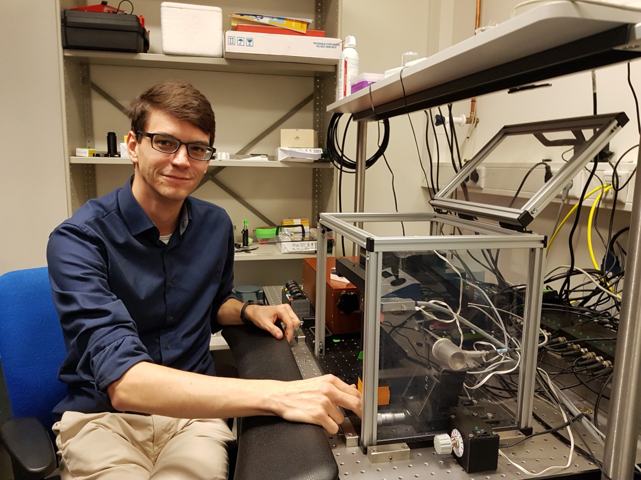
 The awarding of the Nobel Prize to the CRISPR-Cas technique came just at the right moment for Koen Martens. The next day, he defended his (cum laude) thesis, in which the genetic scissors Cas9 play an important role. The microscope he developed, miCube, literally reveals how Cas9 roams along DNA.
The technique in question is called single-particle tracking microscopy, and it follows a single macromolecule. The principle behind the technique has been established for more than five years. Martens improved the technique and applied it to a living cell. He also built a fully open-source microscope to do so, which is already in use in eight places around the world.
Flickering lights
You can follow individual proteins under a microscope by hanging a lamp on them, in this case a flickering fluorescent protein. Martens and his colleagues produced lactic acid bacteria which manufacture dCas9 (Cas without the scissors function) with this lamp. As a result, about 400-500 of these flickering lamps are constantly active in the bacteria. That creates a chaos of flickering spots of light.
So Martens developed a new algorithm that can calculate the location of the lights very precisely. Further calculations can then reveal the route taken by the fluorescent protein in the cell. And it turns out that Cas9 jumps along the DNA to get to where it needs to be. To be precise: it looks for the PAM, a short sequence of code that is indispensable for gene-cutting.
The nice thing is that you can describe the dynamics between Cas9 and the infecting virus
Koen Martens, researcher Biophysics
Martens’ research showed that Cas9 sticks to the PAM for an average of 17 milliseconds before letting go and continuing its search. That happens about 25 times per second. ‘The nice thing is,’ says Martens, ‘that you can describe the dynamics between Cas9 and the infecting virus. You can calculate how long it takes before a single Cas9 finds the virus, or how many Cas9 molecules it takes to eliminate the virus before it multiplies.’
Tracking proteins
‘You can also predict how many CCas9 molecules you need to use to modify a gene,’ says Martens. This kind of microscopy has countless uses. ‘In theory you can keep track of any protein in a living system this way. We couldn’t do that till now.’ Martens now has a postdoc position at the prestigious Carnegie Mellon University in Pittsburgh (Pennsylvania), where he will continue working on applications of this technique.