 Foto: Shutterstock
 Text: Nicole van ‘t Wout Hofland
These structures play a crucial part in the release of flavours. The researchers published their findings in the scientific journal Langmuir.
Foods contain thin, long chains of, for example, glucose or proteins. If these chains form an open network, the flavour is released easily, whereas a more dense network retains the flavour. ‘In the ideal situation, food contains a combination of structures’, says Koen Martens, PhD candidate and lead author of the publication. A network of long, open chains, combined with dense areas, ensures a slow and persistent release of flavours, which makes the taste experience last longer.
Pingpong balls
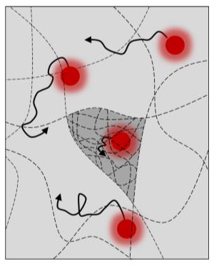
Despite the importance of structures, there was no adequate method to determine the complex organisation of the structures. Thus, Martens and his colleagues developed a method that uses tiny fluorescent spheres. The movement of the tiny balls revealed information on the way the chains are organised. ‘You could compare the system to fluorescent ping-pong balls in a tank of water in a dark room’, Martens says.
To make plant-based meat substitutes appear as meat-like as possible, their protein chains should be similarly arranged
Koen Martens, Biophysics researcher
The balls move through the water freely. But, should you add obstacles to the tank, they can no longer move in random directions. If you study the fluorescent ping-pong balls for a while, it will show you exactly where the balls can move, and thus, where the obstacles are. This is precisely how Martens’ method works, on a much smaller scale: with ping-pong balls of 30 nanometres weaving through an obstacle course of long chains.
Do-it-yourself microscope
‘For us to see these spheres and make the organisation of the structures visible, we needed an extremely sensitive microscope with a powerful laser’, Martens explains. ‘A microscope of that type is not available, and would cost half a million euros to buy’. To economise, the PhD candidate built his own microscope, using aluminium blocks and 3D printed parts.
Using the microscope, the researchers applied the method to the thickening agent carrageenan, a food additive containing long sugar chains. They observed that the sugar chains in the thickening agent were tightly packed in some places, and further apart elsewhere. Thus, they proved that the method is able to identify different chain structures, something that was hitherto impossible.
Meat substitutes
This newly developed method will allow scientists to study the structure of more complex foodstuffs, such as meat substitutes. Meat contains long protein chains, which are arranged in the same direction. ‘To make plant-based meat substitutes appear as meat-like as possible, their protein chains should be similarly arranged’, Martens states. This new method can provide insight and could bring us one step closer to ‘plant-based meat’.
The VLAG Graduate School funded Martens’ and his colleagues’ research.ns weer een stap dichter naar ‘plantaardig vlees’.